Injured koala? Call 1800 775 625 for 24/7 rescue
Koala Chlamydia and Retrovirus Research Project
Overview
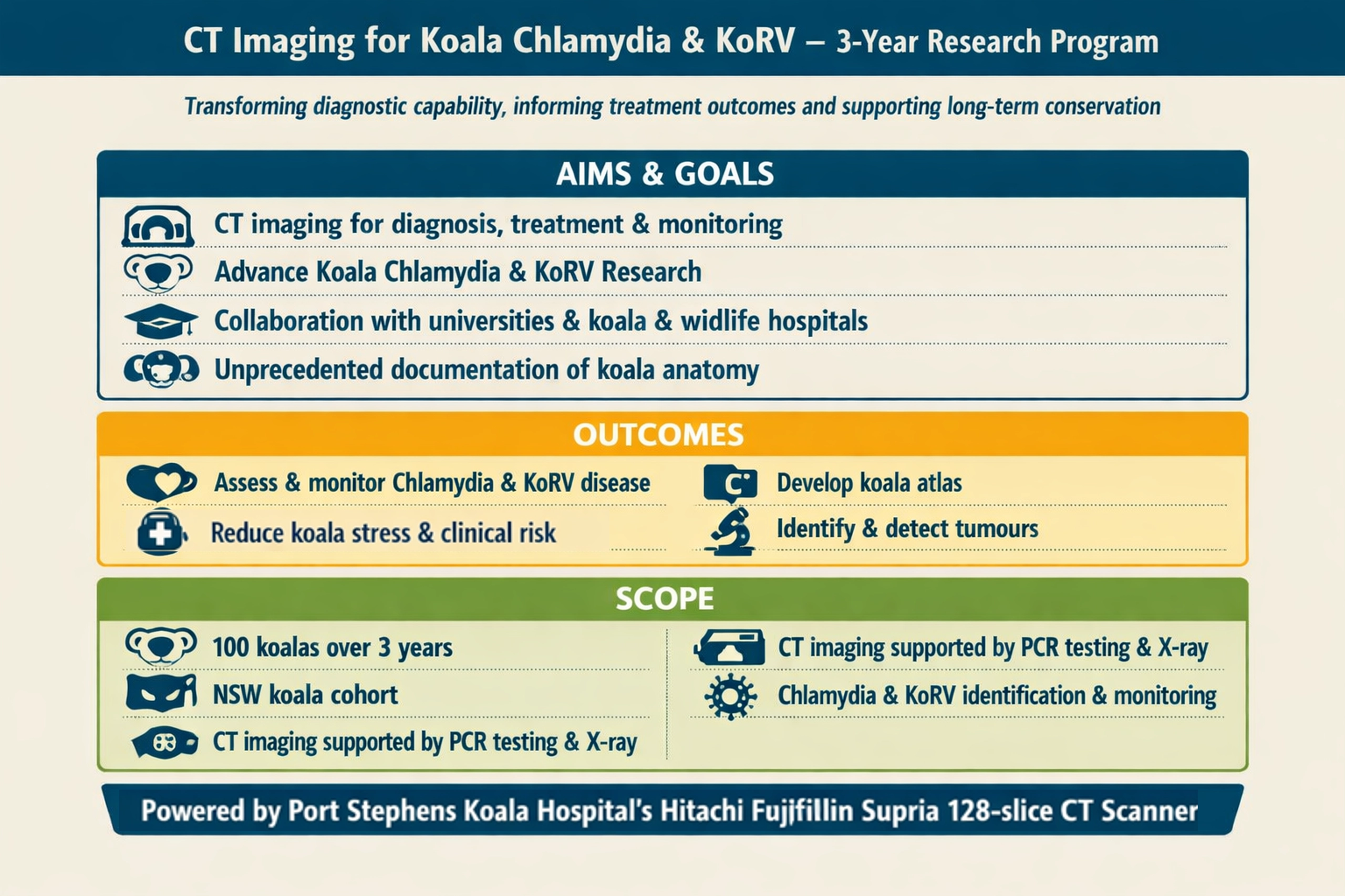
This project uses an advanced computed tomography (CT) scanner to diagnose, treat and monitor chlamydia and koala retrovirus in 150 koalas.
CT imaging is being used alongside established diagnostic techniques such as radiography (X-ray) and ultrasonography to improve diagnostic capability in koala and native wildlife care, supporting more accurate assessment and treatment outcomes.
Current techniques are borrowed from domestic animal medicine and adapted to suit the unique anatomy and physiology of wildlife species. CT will increase the number of studies performed, contributing to our understanding of normal anatomy and disease processes.
Chlamydia
A bacterial infection koalas catch through mating, from mother to joey, or from contaminated environments. It can cause eye disease, urinary issues, and infertility. It’s a different strain to humans.
Koala retrovirus (KoRV)
A virus that’s become part of koala DNA – many are born with it. It spreads from mother to offspring and can weaken the immune system, making koalas more vulnerable to chlamydia. Humans can’t catch it.
Research funded by the Australian Government

Onsite project upgrades
To support the project, we’ve put a number of key infrastructure and facility enhancements in place across the hospital.
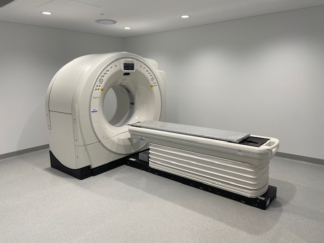
- Installation of a Hitachi Fujifilm Supria 128-slice CT scanner
- Generator installation to support CT operations
- Koala holding and quarantine enclosures
- Bio-Rad CFX Opus 96 qPCR system for on-site chlamydia testing
- High end imaging hardware and software imaging management
- Purchase of a multi-purpose rescue/release off-road vehicle (Milestone 3, from July 2027)
The project is supported by specialist veterinarians, radiographers, veterinary nurses, wildlife rehabilitators and trained volunteers – all working to the highest clinical and ethical standards.
Standard diagnostic techniques
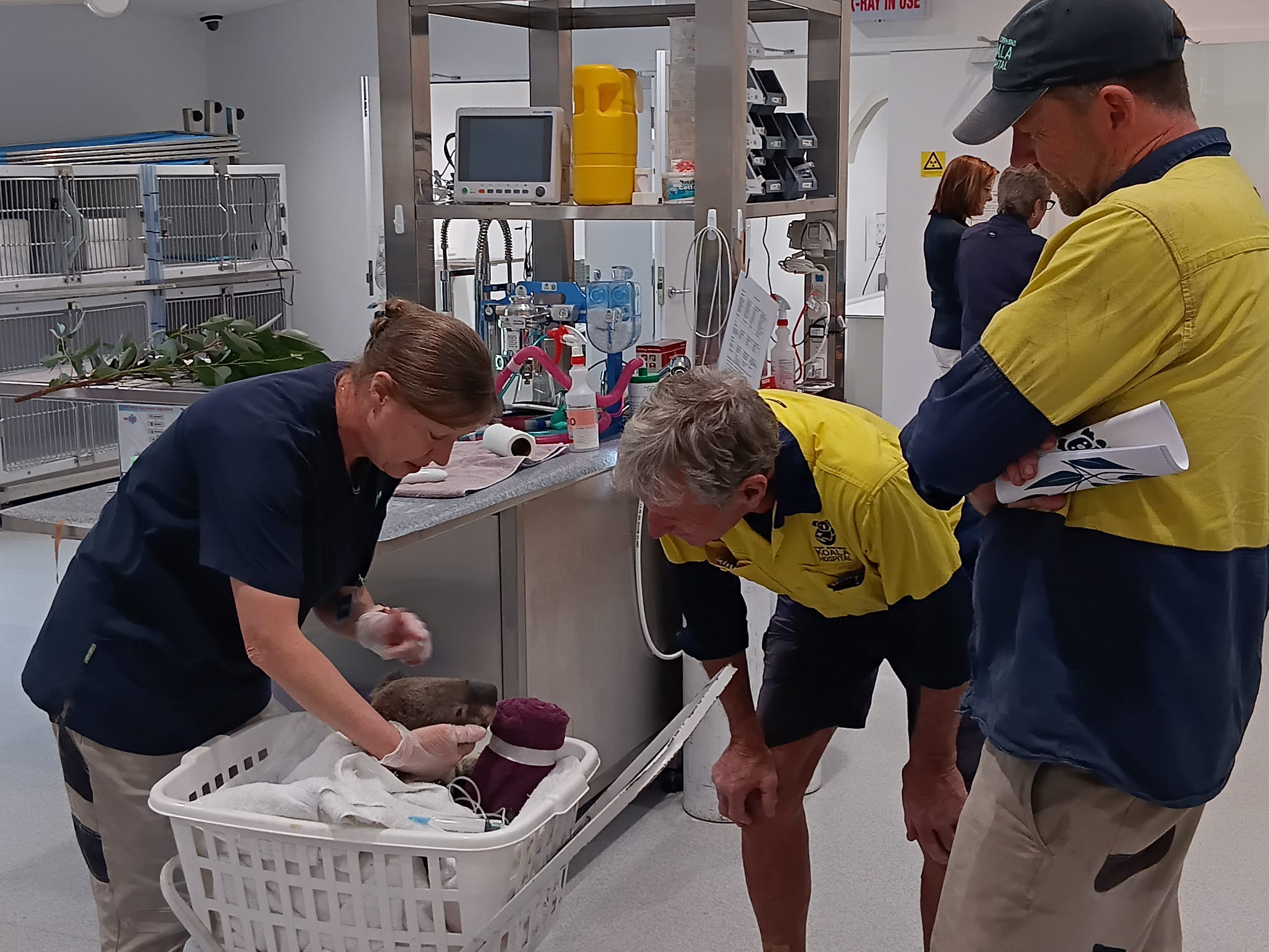
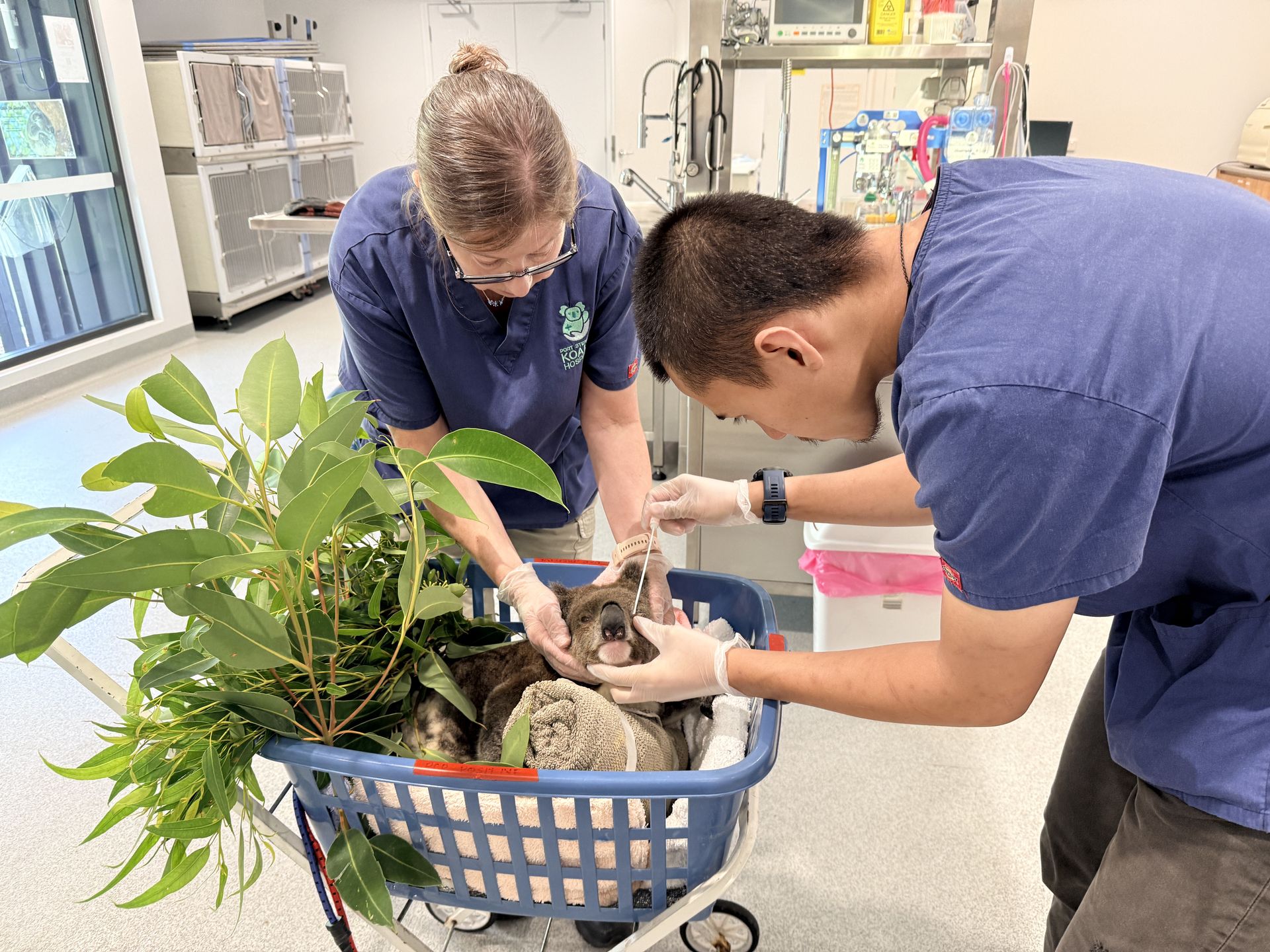
All koalas in our care undergo routine diagnostic testing on admission. This involves extensive exams and testing to determine what injuries they have and to look for signs of disease.
- Physical exam with a vet
- Blood taken for biochemistry and haematology
- Swabs taken of multiple sites to test for chlamydial DNA
- Ultrasound of the koala, looking especially at the reproductive and urogenital tract
- Radiographs of the koala looking at the skeleton, chest, and abdomen.
- Mucosal swabs and hair samples are taken for the Australian Museum DNA database.
Advanced CT scanning
Hitachi Fujifilm Supria 128-slice
This machine captures rapid, whole-body 3D scans of koalas in fine detail, building images of all essential organs, including kidneys, uterus, ovaries, bladder, using up to 128 image slices. It allows koalas to be imaged inside their carrying containers, without handling or sedation.
- Assess and monitor disease and injury more safely
- Enables unprecedented documentation of koala anatomy
- Improves accuracy of diagnosis and prognosis
- Lowers koala stress and risk from sedation and anaesthesia
- Reduces repeat interventions, improving survival rates
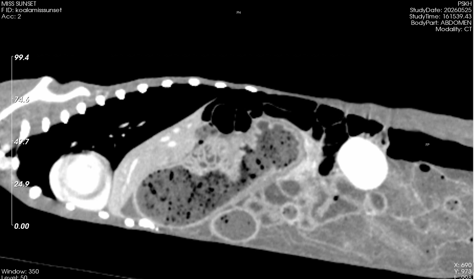
Baseline assessment
10 koalas – both live and deceased – underwent comprehensive health checks and medical examinations, including ultrasound, biochemistry, haematology, radiography, physical examination and CT scanning.
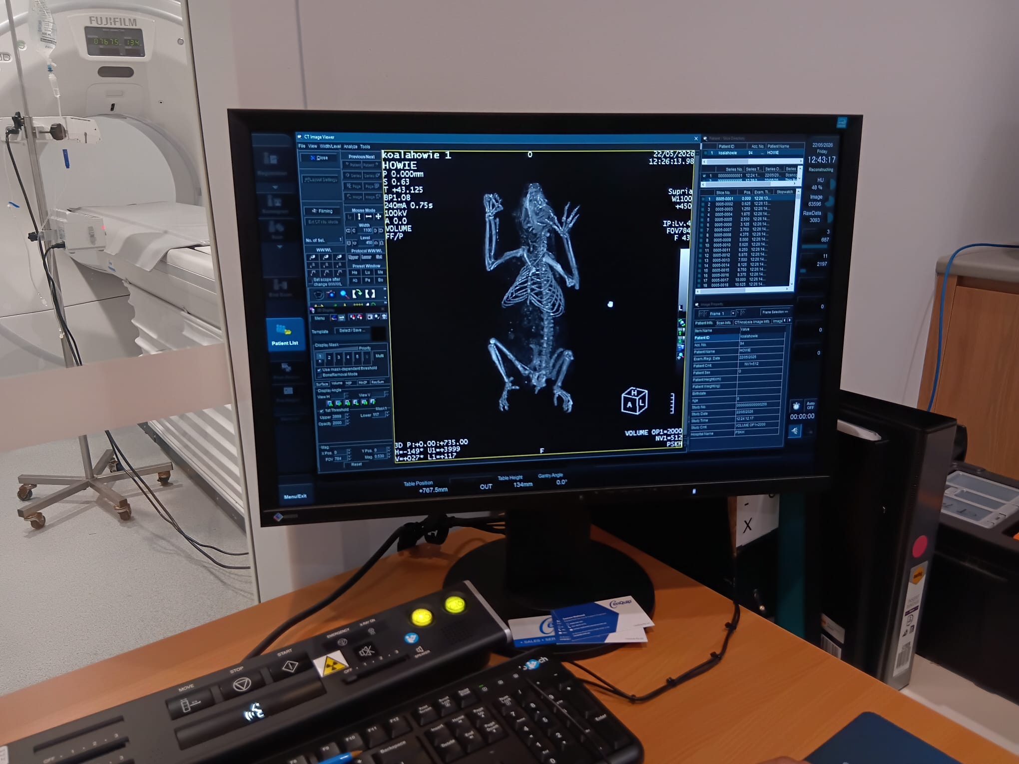
Research methodology
Step 1 – Collection: Each koala is identified and collected for examination.
Step 2 – Examination: Each koala undergoes a full clinical examination, including sedation, sampling, diagnostic testing and CT scanning. Key areas of examination include the urogenital tract, nasal structure, respiratory tract, musculoskeletal system and lymphatic system.
Step 3 – Testing: CT findings are compared against ultrasound, x-ray and postmortem results to better understand how chlamydia develops, spreads and resolves. Chlamydia testing is conducted onsite using the Bio-Rad CFX Opus 96 qPCR system.
Step 4 – Recovery: Each koala is monitored for 1–2 hours before being safely returned to their enclosure.
Step 5 – Analysis: Our specialist team reviews all results, including radiographs, CT data and haematology.
Step 6 – Confirmation: A selected number of koalas will undergo postmortem at Sydney University to validate imaging findings.
The new qPCR testing system enhances detection of chlamydia providing more accurate and faster results than previous methods.
Expected research outcomes
- Assess and monitor Koala Chlamydia and Koala Retrovirus disease
- Improved long-term health and survival rates for koalas in care
- Creation of a 3D koala anatomy atlas, building on the existing Augmented Reality Program from the Save Koala Fund Project
- Greater understanding of normal koala anatomy as a reference for future treatment
- Greater understanding of normal and diseased koala anatomy to inform future treatments
- A replicable model for koala and native wildlife research across Australia